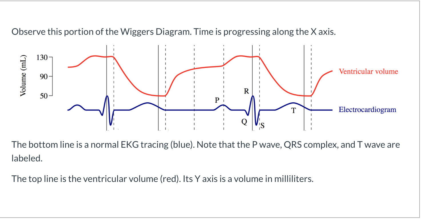
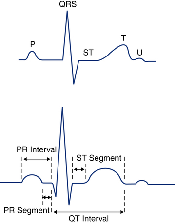
The Portion of the Tracing of an Electrocardiogram
The QRS complex one single heart beat corresponds to the depolarization of the right and left ventricles lower heart chambers. The word ECG derives from the German language.

Typical Lead Ii Electrocardiographic Tracing Showing Standard Download Scientific Diagram
There are some basic rules that can be followed to identify a patients heart rhythm.

. Typically the isoelectric line is measured as the portion of the tracing following the T wave and preceding the next P wave. Part B-Electrocardiogram Tracing. Routine electrocardiogram of the heart EKG with tracing using at least 12 leads.
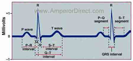
This tracing consists of representations of several waves that recur with each heartbeat about 60 to 100 times per minute. Five small squares equal 020 seconds. An ECG electrode is a conductive pad that is attached to the skin to record electrical activity.
This isoelectric period represents a time period during which the ventricles are completely depolarized plateau phase. Segment on the EKG tracing may vary from that above depending on the lead used or the presence of abnormal conduction. Atrial repolarization occurs during this time as well.
The procedure for producing an electrocardiogram. The QRS complex represents ventricular depolarization and contraction. The T wave represents the re-polarization or recovery of the.
The portion of the tracing of an electrocardiogram ECG that represents ventricular depolarization is the _____. Conversion table for heart Rate. Drag and drop the correct labels to the appropriate location on this ECG Reset Help repolarzation P wave aa Incorrect.
It indicates that the atria are contracting pumping blood into the ventricles. Is the first short upward movement of the EKG tracing. The data is recorded to a paper printout sometimes called a tracing.
The horizontal lines measure time intervals and heart rate. Anatomy and Physiology questions and answers. This event consists of a number of health care services that often occur at the same time.
The hearts electrical waveform can be detected by electrodes placed on the surface of the body. Our new EKG modules provide in-depth training in EKG basics rhythm analysis and a wide range of EKG topics. T wave QRS complex Q wave.
An EKG is a representation of the electrical activity of the heart muscle as it changes with time usually printed on paper for easier analysis. Does not include interpretation or report. What is the rate.
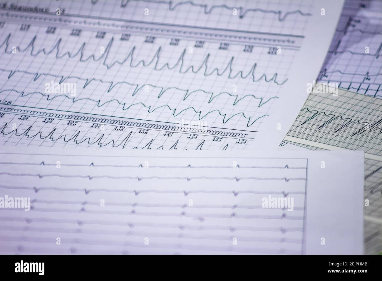
The meaning of ELECTROCARDIOGRAM is the tracing made by an electrocardiograph. An ECG detects your hearts electrical rhythm and produces whats known as a tracing which looks like squiggly lines. These 3 second time intervals are marked on the paper by darker lines as shown below.
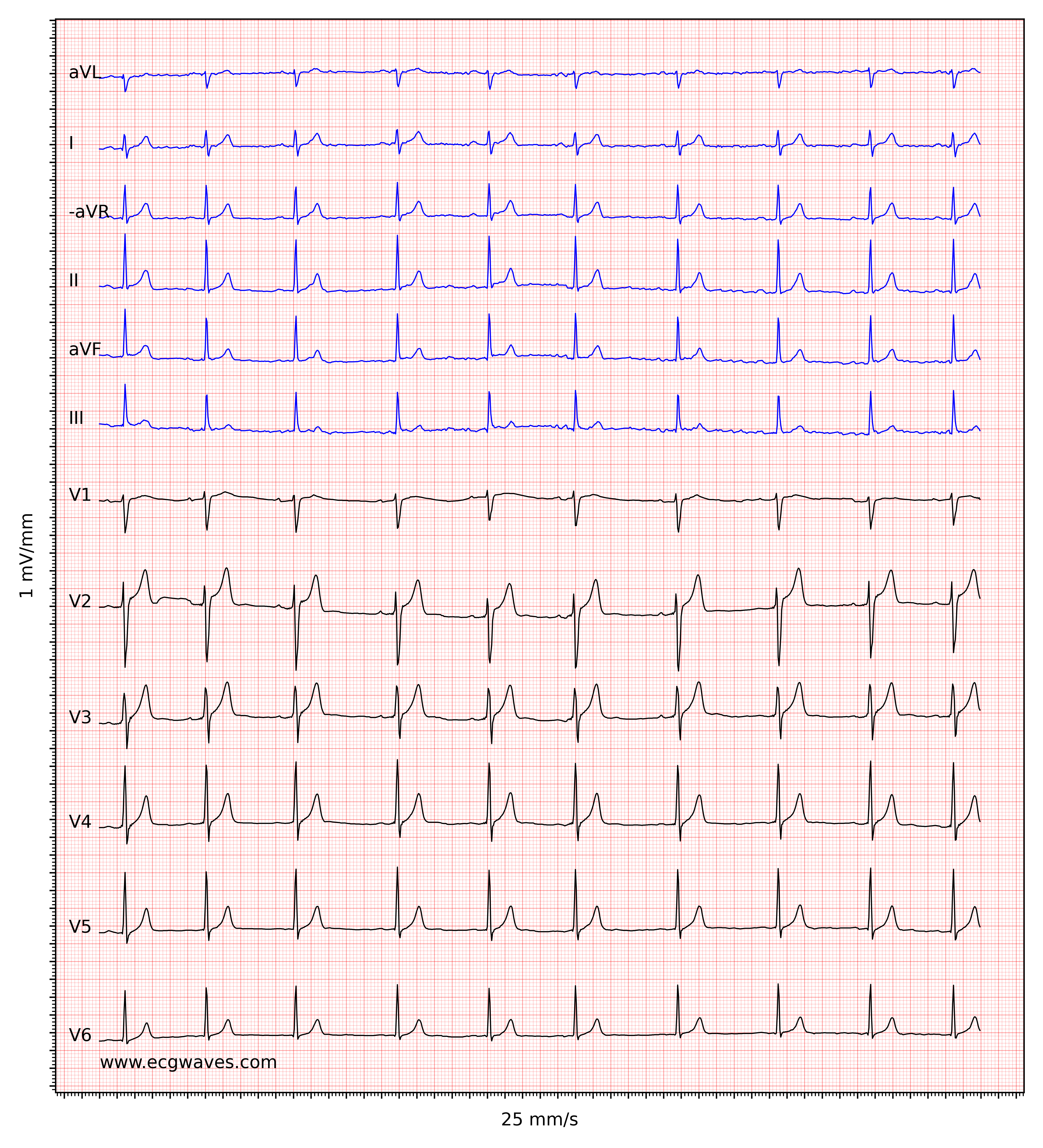
An ECG lead is a graphical representation of the hearts electrical activity which is calculated by analysing data from several ECG electrodes. Each of the small squares equal 04 second of time. P Q R S T corresponding to different phases of the heart activities.
The electrocardiogram abbreviated as ECG or EKG represents an electrical tracing of the heart and is recorded non-invasively from the surface of the body. The portion of the tracing of an electrocardiogram ECG that represents ventricular depolarization is the _____. Up to 10 cash back Find the perfect ecg heart tracing stock photo.
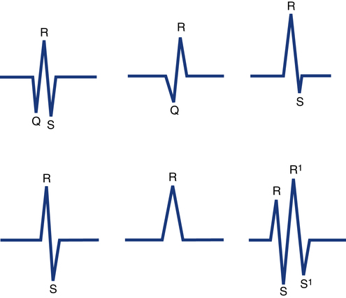
Often the Q portion is not evident and the depolarization presents as only an RS complex. The electrocardiogram EKG is a graphic representation of the electrical activity of the heart. This is because the ECG does not directly depict abnormalities in cardiac structure such as defects in the heart valves or interventricular septum.
As the cells repolarize in this area the cells gain positive charges on the outsides of the membrane and a dipole or separation of charge across the heart surface can be measured again. Normally beginning with a downward deflection Q a larger upwards deflection R. QRS complex is the portion of the EKG tracing that represents depolarization in the myocardium and the ventricle are activated simultaneously.
The P wave represents the normal atrium upper heart chambers depolarization. The baseline voltage of the electrocardiogram is known as the isoelectric line. Each lead of the EKG looks at a specific part of the heart from different angles.
A 12-lead ECG records 12 leads producing 12 separate graphs on a piece of ECG paper. From the initial 3 leads of Einthoven who inaugurated electrocardiography current EKGs are composed of 12 leads each of which records the electrical pattern from a slightly. And then a downwards S wave.
EKGs can be used to diagnose heart attacks heart problems including electrical malfunctioning and other heart problems. Fifteen of the 020 squares represent 3 seconds. The EKG is a printed capture of a brief moment in time.
In 1902 the Dutch physician Einthovan invented ECG and. It is important to note that the ECG tracing does not measure the pumping ability of the heart. Electrocardiogram ECG or EKG With Tracing.
ECG tracing waves explained Images by JS BYU-Idaho F17 Finally the cells of the apex begin to repolarize. Use the button below to learn more. It is not unusual for a patient with a low cardiac output to have a normal ECG tracing.
No need to register buy now. Another limitation worth noting is that. T-wave is deflection of the normal electrocardiogram following QRs complex and it represent repolarization or recovery of the ventricle.
When your childs doctor studies the tracing of the EKG they look at the size shape and length of the waveforms and how fast or slow the heart is beating. Each ECG cycles consists of 5 waves. The wave pattern should have a consistent shape.
Each module includes lessons interactive drills and a course quiz. For people who are 50 or older getting an ECG as part of an annual physical exam makes sense some cardiologists say. An electrocardiogram ECG is a quick painless noninvasive test that can help diagnose dozens of heart conditions.
The ECG coach breaks down interpretation into five analysis steps followed by classification of the ECG tracing. Part B-Electrocardiogram Tracing. The Q portion is the initial downward deflection the R portion is the initial upward deflection and the S portion is the return to the baseline or the so-called isoelectric point.
In German it is elektro-kardiographie. Is it regular or. QRS complex Which heart chamber receives blood from the lungs.
Huge collection amazing choice 100 million high quality affordable RF and RM images.

Ecg Basics Ppt Video Online Download

Electrocardiogram Ecg Tracings Depict Prospective Ecg Triggering And Download Scientific Diagram
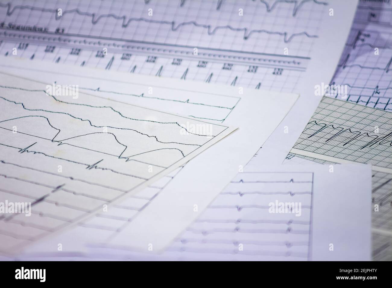
Tracing Of Electrocardiograms On Graph Paper Records Of Heart Activity Heartbeat With Arrhythmia Selective Focus With A Portion Out Of Focus Stock Photo Alamy
Solved Question 1 Recall When Are The Atria In The Process Chegg Com
Ecg Interpretation Characteristics Of The Normal Ecg P Wave Qrs Complex St Segment T Wave Ecg Echo

Electrocardiogram Ecg Tracings Depict Prospective Ecg Triggering And Download Scientific Diagram

Pressure Waveform Pitfalls And Abnormalities Ca Cannon A Wave Cvp Download Scientific Diagram Central Venous Pressure Abnormal Pressure
Interpretation Of Electrocardiogram Tracings Clinical Gate

What Is An Ecg

A Typical One Cycle Ecg Tracing 1 Download Scientific Diagram
Interpretation Of Electrocardiogram Tracings Clinical Gate

The Electrocardiogram Thoracic Key

The Physiological Measurement Handbook
Tracing Of Electrocardiograms On Graph Paper Records Of Heart Activity Heartbeat With Arrhythmia Selective Focus With A Portion Out Of Focus Stock Photo Alamy
Ecg Ekg Components

A Typical Ecg Tracing Of The Cardiac Cycle Pdf Electrocardiography Cardiac Electrophysiology
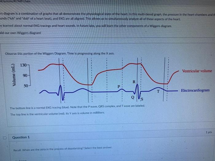
Solved 4 Quizzes 4074481 Take Es Diagram Is A Combination Of Chegg Com

Electrocardiogram From A 19 Year Old Woman After Near Drowning A Download Scientific Diagram
And Shape Of Electrocardiogram Veterian Key


Comments
Post a Comment